


The medical scholars during the medieval Islamic era placed great emphasis on the value of dissection and the knowledge of anatomy for the diagnosis of affected organs, the relationships of the organs to one another and the application of adequate medical and surgical treatment.

***
Note of the Editor: We are pleased to announce the publication of Dr Rabie E. Abdel-Halim’s “1001 Cures: Introduction to the History of Islamic Medicine” as the second in the series of 1001 Cures books with this article “Human Anatomy” from Chapter Four, Pages 85-89, published by the Foundation for Science Technology and Civilisation (FSTC).
***

Figure 1: Avicenna depicted in the Daqa’iq al-Haqa’iq by Nasir ad-Din Rammal in the 14th century CE (Source)
Thus, they emphasised that the doctor should be quite knowledgeable in anatomy. Hence, Abū ʿAlī al-Ḥusayn ibn ʿAbd-Allāh Ibn Sīnā (Avicenna, 980–1037) made it clear that, if the medical treatment for a bladder stone fails and the cutting operation had to be carried out, one must choose:
“the one who knows the dissection of the bladder, the places where it is joined at its neck by the semen channels, and the related vessels so that he could prevent what he should keep away from, such as causing an inability to reproduce or heavy loss of blood or a fistula that does not heal”
Also Abū al-Qāsim Khalaf ibn al-ʿAbbās al-Zahrāwī (Albucasis, 936–1013) in the thirtieth maqalah of his encyclopaedic work al- Tasrīf, devoted to surgery and surgical instruments stated that:
“He who is not skilled in as much anatomy as we have mentioned is bound to fall into an error that kills a human being.”
Similarly, Abū Marwān ʿAbd al-Malik ibn Abī al-ʿAlāʼ ibn Zuhr (Avenzoar, 1093– 1162) emphasised the great importance of practical knowledge of dissection in the following warning in his book al-Taysīr, in the course of discussing the management of inflammatory swellings of the neck when ripe and ready for bursting or drainage:
“And in case you have mastered the science of dissection, then drain by the scalpel in the way that you will not come across a vein, artery, nerve or anything whose injury will lead to an extra harm to the patient”
In addition, Alāʾ al-Dīn Abū al-Ḥasan Alī ibn Abī al-Ḥazm al- Qarshī ibn al-Nafīs (1210–1288) allocated a special chapter in his book, Sharḥ Tashrīḥ al-Qānūn, entitled On the Benefits of Studying the Science of Anatomy, and showed how essential this study is for reaching diagnoses and for practicing medicine and performing different surgical, orthopaedic or ophthalmological procedures. Furthermore, in this book he wrote a special chapter on the best mode for dissecting the following parts: bones, peripheral vessels and internal organs of the chest (heart, lung, big vessels and the diaphragm).
In fact, all the eminent Islamic physicians and the theologians of this era stated that knowledge of anatomy leads to a deeper appreciation of God’s wisdom and omniscience. In this context, ʾAbū al-Walīd Muḥammad ibn ʾAḥmad ibn Rushd (Averroes, 1125– 1198), a reputed philosopher and medical scholar who was the Grand Qāḍi (Chief Magistrate) of Cordova and a well-known authority on Islamic jurisprudence in the whole Muslim world then, and up until now, stated that “man ishtaghala bi ʿilm al tashrīḥ izdāda ‘imānan billāh” (“anyone who practices the science of dissection will increase [his] faith in God”). Moreover, the word ishtaghala in this statement also has significance and special connotations, as it means “practice” or “become occupied with” rather than denoting a simple theoretical knowledge of anatomy. In confirmation of this, Abū Bakr Muḥammad ibn Zakariyyā al-Rāzī (Rhazes, 854–925), in his book al-Manṣūrī, describes compounds to preserve cadavers. Thus, the practice of dissection for medical teaching in the Muslim world was not prohibited by the religion of Islam.
Ibn Zuhr described two forms of serious and fatal diseases that may affect the heart. One of them he described as follows:
“In the heart, a watery fluid collection looking like urine, may occur. It is found enclosed and contained within its covering”.
For the other disease, he described “solid substances accumulating on the inside of the heart’s covering looking like layers upon layers of membranes”. It seems from these two descriptions that Ibn Zuhr was, most probably, describing the morbid changes in the serous and fibrinous forms of pericarditis. Thus, postmortem dissection to identify the cause and course of fatal diseases seems to have been known and acknowledged in his time. In confirmation of this, Ibn Zuhr, in his search for treatment for ulcerative diseases of the lungs, stated that “remedies for lung ulcerations are around in nature but are yet unknown to us, as sheep, when caught with a lung disease, do leave the herd, as shepherds say, and wander about as if looking for a plant to eat, and when they finish eating it their illness is relieved completely and they are back to normal”. Then, to confirm its cure, Ibn Zuhr said:
“I did inspect lungs of sheep with the evident effect of breach of continuity and with obvious evidence of healing and union”.
Hence, he resorted to postmortem examination of sheep in order to confirm his observation, although he could not identify the herb.

Figure 2: A depiction of Ibn Zuhr from “Arabic Medicine” (c. 1906), by Veloso Salgado (Source)
The medical scholars of the medieval Islamic era, particularly from the time of al-Rāzī onwards, critically appraised the views of those who came before them in the light of their own experience, experimentation and direct observations from their practice of human dissection. This led to the correction of some ancient anatomical data, as will be shown in the following examples.
Contrary to Galen, al-Rāzī’s descriptions of the sacrum (ʿaẓm al-ʿajuz) and coccyx (ʿaẓm al-ʿuṣʿuṣ) are more accurate, as he described the sacrum as consisting of only three parts and stated that they look like vertebrae. Also, he described the coccyx as a separate bone, similarly consisting of three parts, with only the last part looking like a cartilaginous bone. Furthermore, Ibn Sīnā described the parts of the sacrum as “vertebrae with the strongest construction, most tight joints and widest wings”. Meanwhile, he recognised the independent nature of the coccyx as separate from the sacrum and, unlike al-Razi, he described it as consisting of three cartilaginous bones. Therefore, later Muslim scholars such as al-Zahrāwī, Amīn al- Dawlah Abū al-Faraj ibn Yaʿqūb ibn Isḥāq Ibn al-Quff (1233–1286), and Ibn al-Nafīs directly included the sacral and coccygeal vertebrae in the total count of the bones of the spine making them a total of thirty.
Galen’s concept of the lateral movement of the head in both directions and forward and backward movements (nodding), although it had been accepted by ʿAlī ibn al- ʿAbbās (Haly Abbas, d. 982–994), Ibn Sīnā and other Muslim scholars, was criticised by Ibn al-Nafīs in his commentary on the anatomy of Ibn Sīnā’s Canon of Medicine.

Figure 3: Galen dissecting a monkey, as imagined by Veloso Salgado in 1906 (Source)
Furthermore, contrary to Galen’s description of the body of the lower jaw, the 12th-century Muwaffaq al- Dīn ʿAbd al-Laṭīf al-Baghdādī (1162–1231) confirmed the unitary nature of the lower jaw by experimentation in order to check the validity of his predecessor’s knowledge. The detailed description of this pioneering anatomical experiment is hereby translated from his book Kitāb al-ʿIfādah wa-al-‘Iʿtibār ( Book of Utility and Verification):
“…with regard to the shape, proportion and relations of bones and joints in a large number of the examined human skeletons, we have gained knowledge that we could not obtain from books, either because of being overlooked or because of lack of textual clarity or because our findings are different from what is written in those books, though indeed, direct observation is stronger evidence than hearing. Although Galen possessed the highest qualities in checking and verifying what he reports, direct observation is a more true source of knowledge than his reports. Then, an attempt can be made, if possible, to think of an explanation for his views. Among those discrepancies is the lower jaw bone, which all agreed with Galen to describe as consisting of two separate bones joined at the chin by a strong joint… However, based on our own observations, this organ (mandible) is, first and foremost, one bone only without a joint or a symphysis. Using various methods of testing, we examined it repeatedly as many times – as willed by Allah – in many specimens whose number exceeded two thousand skulls; but from all aspects, we did not find it except as a single bone. We also arranged the assistance of a distinct group, who examined it (the mandible) in our presence and then in our absence. They did not add anything to our observations and reporting.” (See Figure 4)
Thus, the large number of the examined specimens also reflects his awareness of the statistical importance of the sample size in determining the significance of findings. Moreover, in order to avoid any possibility of bias, Muwaffaq al-Dīn al-Baghdādi repeated his experiment three times; first on his own, then together with a group of scholars and finally through another group of scholars on their own, so, to achieve accuracy of results, he utilised more than one method of testing.

Figure 4: Part of page 61 of Muwaffaq al-Dīn ʿAbd al-Laṭīf al-Baghdādī’s book Kitāb al-‘Ifādah waal-‘ Iʿtibār (Book of Utility and Reflection) containing the report on his anatomical study of more than 2000 human skulls.
Likewise, Ibn Sīnā, in the course of his study of moisture in various organs of the body, including bones, described the following experiment:
“Take two equal-weight amounts of bone and hair, then distil them in the alembic. This results in more moisture and fat leftover from the bones with a less weight residue than that of the hair”. Thus, he concluded that “bones are more humid than hair”
Contrary to Galen, Ibn Zuhr permitted a form of sensation for both bones and teeth, though of a better kind in the latter. He stated that as bones, in a living body, grow and do not decay, although there is no hairlike branching of veins and arteries in them, so, they can, similarly, have sensation, although the nerve does not branch like hairs inside them. Ibn Zuhr then said that:
“This is what I thought based on analogy and logical deduction”.
He pointed out that the real answer to this matter is to verify it by dissection, which he did not do.
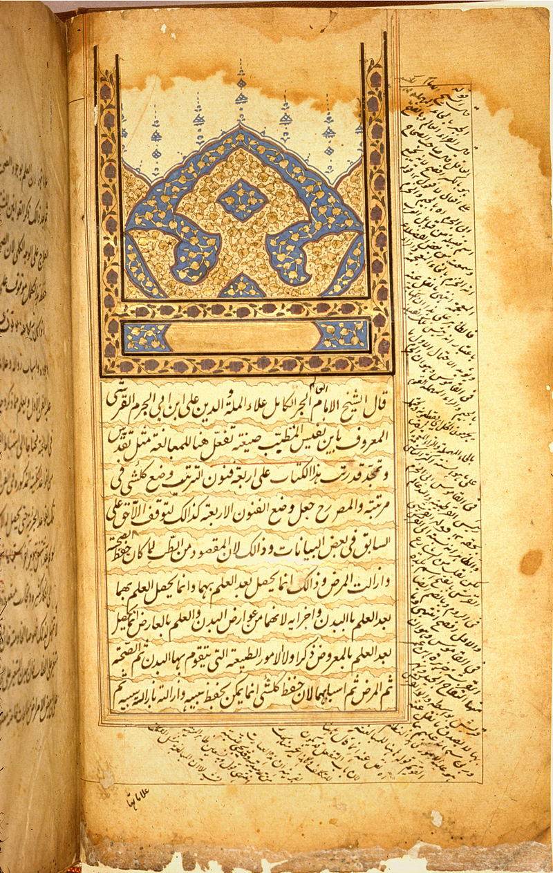
Figure 5: Opening page of one of Ibn al-Nafis’ medical works. This is an Indian during the 17/18th century (Source)
Though Ibn Sīnā stated that the heart is located in the centre of the thorax and slightly tilted to the left, all other scholars, contrary to Galen, stated that, though the heart is in the centre of the thoracic cavity, its apex is directed to the left side. Ibn Sīnā, as well as all the scholars, agreed with Galen that the heart has two ventricles: the right containing thick blood, and the left containing thin blood. However, in addition, Ibn Sīnā described the third ventricle as a storehouse for the nutriments of the heart, which was refuted by Ibn al-Nafīs. In the medieval Islamic era, all the scholars before Ibn al-Nafīs, namely, al-Rāzī, ʿAlī ibn al-ʿAbbās, al-Zahrāwī and Ibn Rushd, agreed with Galen’s statement that the two ventricles of the heart are communicated through the interventricular septum by invisible pores. However, Ibn al-Nafīs stated that:
“There is no connecting passage in between the two cavities because the heart substance there is compact without any obvious passage, as thought by some, or invisible passage that could transmit that blood as thought by Galen. Indeed the texture of the heart there is compact and its substance thick”
Contrary to Galen, ʿAlī ibn al-ʿAbbās confined the pulsations only to the arteries and introduced the names “al-ʿUrūq al-Ḍawārib” (pulsating vessels) for the arteries and “al-ʿUrūq Ghair al-Ḍawārib” (non-pulsating vessels) for the veins, thus highlighting the difference between arteries and veins.6With the name “al-ʿUrūq Ghair al-Ḍawārib”, sometimes replaced by “al-ʿUrūq al-Sawākin (the motionless vessels), this nomenclature continued in the works of most of the scholars who came after ʿAli ibn al-ʿAbbās.
Also unlike Galen, all the scholars al-Rāzī, ʿAlī ibn al-ʿAbbās, al-Zahrāwī, Ibn Sīnā, Ibn Rushd and Ibn al-Nafīs described the arteries to the heart in more detail. They stated that two arteries emerge from the aorta at its beginning, the large one circulates around the head of the heart along with the vein and divides into branches which penetrate its substance, whereas the smaller one turns right, penetrates and divides within the substance of the right ventricle.
Furthermore, Ibn al-Nāfīs, in his description of the attachment of the artery-like veins (pulmonary veins) to the heart, stated that:
“The growing out of these two artery-like veins (pulmonary veins) is not from the left ventricle al-tajwīf al-aysar but from the structure ‘al-jurm’ that lies between the two ventricles of the heart, though they are inclined towards the left ventricle so that their cavities connect obliquely, with its cavity as if the direction of outlet of that cavity is slightly deflected to the right so that it continues with their lumens”.
Thus, his description may, possibly, denote an early description of the left atrium.

Discover the golden
age of Muslim civilisation.
© Copyright FSTC Ltd 2002-2020. All Rights Reserved.


_-_Veloso_Salgado.png)