Al-Razi (Rhazes) (born in 864 CE) wrote over 200 scientific treatises, many of which had a major impact on European medicine. His best known manuscript is Liber Continens, a medical encyclopedia in which he described his contributions to neurology, focusing on his description of cranial and spinal cord nerves and his clinical case reports, which illustrate his use of neuroanatomy to localize lesions. In this article, Dr Nizar Souayah and Dr Jeffrey I. Greenstein focus on Al-Razi's description of the cranial and spinal nerves and his relevant clinical case reports, which illustrate his understanding of neuroanatomy and the application of his knowledge to clinical practice.

Dr Nizar Souayah, MD; and Dr Jeffrey I. Greenstein, MD*
Table of contents
1. Abstract
2. Background
3. Methods
4. Results and discussion
5. Conclusion
6. Appendix
7. Footnotes and references
***
Note of the editor
This article was first published as Dr Nizar Souayah, MD; and Dr Jeffrey I. Greenstein, “Insights into Neurologic Localization by Rhazes, a Medieval Islamic Physician”, Neurology 2005;65;125-128 (online at https://www.neurology.org/cgi/content/full/65/1/125). Print ISSN: 0028-3878. Online ISSN: 1526-632X. Copyright © 2005 by AAN Enterprises, Inc. We thank Dr Nizar Souayah for granting us the right to republish it.
***
1. Abstract
Rhazes was born at Ray near modern Tehran in 864 CE. He wrote over 200 scientific treatises, many of which had a major impact on European medicine. His best known manuscript was Liber Continens, a medical encyclopedia. Herein are described Rhazes’s contributions to neurology, focusing on his description of cranial and spinal cord nerves and his clinical case reports, which illustrate his use of neuroanatomy to localize lesions. Relevant passages from facsimiles of the manuscripts Kitab al-Hawi (Liber Continens) and Al-Mansuri Fi At-Tibb (Liber Al Mansoori) were translated, reviewed, and used as references. In addition, Medline, Web, and manuscript searches on Rhazes and the history of medieval and Islamic medicine and neurology were conducted. Rhazes stated that nerves had motor or sensory functions, describing 7 cranial and 31 spinal cord nerves. He assigned a numerical order to the cranial nerves from the optic to the hypoglossal nerves. He classified the spinal nerves into 8 cervical, 12 thoracic, 5 lumbar, 3 sacral, and 3 coccygeal nerves. Rhazes showed an outstanding clinical ability to localize lesions, prognosticate, and describe therapeutic options and reported clinical observations, emphasizing the link between the anatomic location of a lesion and the clinical signs. Rhazes was a pioneer in applied neuroanatomy. He combined a knowledge of cranial and spinal cord nerve anatomy with an insightful use of clinical information to localize lesions in the nervous system.
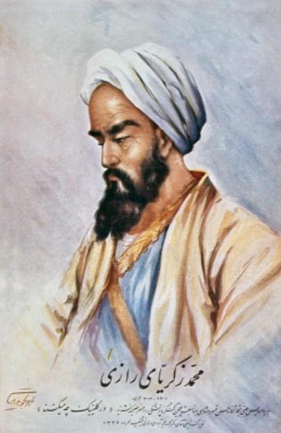
Figure 1: Imaginary portrait of al-Razi by a modern artist. (Source).
The correlation between neuroanatomy and clinical information was pioneered by the Greeks and Romans, Galen (131 to 201 CE) in particular. In the Middle Ages, Rhazes (864 to 930 CE) played a major role in recognizing and describing lost ancient contributions to this clinical skill. In addition, he made his own contributions at a time when the art of clinical medicine in Europe was dormant. Here we report the descriptions of cranial and spinal nerves by Rhazes and his use of neuroanatomy to localize neurologic lesions.
2. Background
Abu Bakr Muhamed Ibn Zakaria Al-Rhazi was born at Ray in Khurasan, near modern Tehran, in 864 CE. Early in his life, he was interested in music and arts. Later on, he became interested in philosophy, mathematics, astronomy, and chemistry. He studied medicine in his 30s or 40s and completed his medical training in Muqtadiri Hospital (the main hospital of Baghdad), mainly under Ali Ibn Rabane (died 933 CE). He also studied under the supervision of a student of Ibn Isaac, a physician well versed in Persian, Greek, and Indian medical systems. After finishing his training, Rhazes left Baghdad for Ray, his native city, to take charge of the local hospital, where he gained eminence as a clinician. Students and patients flocked to him from distant parts of the Islamic empire. Because of his reputation, the Caliph in Baghdad appointed him the head of the main hospital in Baghdad and a court physician in 907 CE. After many years in Baghdad, he returned to Ray, where he died in 930 CE [1].

Figure 2: The final page of the copy of the Hawi by al-Razi, with the colophon in which the unnamed scribe gives the date he completed the copy as Friday, the 19th of Dhu al-Qa`dah in the year 487 (= 30 November 1094). The National Library of Medicine, Bethesda, Maryland, MS A17, p. 463. This manuscript is the third oldest Arabic medical manuscript known to be preserved today. (Source).
Rhazes is considered one of the two greatest physicians in medieval medicine. While the other, Abu-‘Ali al-Husayn ibn-‘Abd-Allah ibn-Sina (Avicenna, 937 to 1037 CE), contributed to the theory of medicine, Rhazes contributed to the practice of medicine [2]. He is regarded as one of “the most original of the Arabic writers who followed both Hippocrates and Galen in their methods and their ideas [3].” Rhazes combined knowledge of textual scholarship with astute clinical observation [4]. He left 200 scientific treatises on various subjects [5]. The most well known is Kitab Al Hawi Fi Al-Tibb, also known as Liber Continens, a 25-volume medical encyclopedia [6]. Here he translated some work of Greek physicians and added material from other Greek as well as Indian and Arabic authors, interspersing this with knowledge from his own experience [7]. Liber Continens was one of only nine volumes in the library of the Paris Faculty of Medicine in 1395 CE and was among the most widely read medieval medical manuals in Europe. In another treatise, Kitab Al Mansoori [8], dedicated to al-Mansur, the prince of Khurasan and also known as Liber Al Mansooris, Rhazes dealt with Greco-Arab medicine and devoted an entire chapter to anatomy, including neuroanatomy.
Despite the fact that he was a Galenist in his theories, he followed the principles of Hippocrates in observing his patients, taking a detailed medical history and writing keen notes. Rhazes was a pioneer in many areas of medicine including paediatrics [9], infectious disease [10], neurosurgery [11], ophthalmology [12], and therapeutics [13]. For instance, he wrote the first manuscript on pediatrics, Diseases in Children [14]. He also first described measles as a clinical entity and differentiated it from smallpox in Liber de Variolis et Morbillis [15]. Relatively little is known of his prescient contributions to neurology [16].
In this article, we focus on Rhazes’s description of the cranial and spinal nerves and his relevant clinical case reports; which illustrate his understanding of neuroanatomy and the application of his knowledge to clinical practice.


Figure 3 a-c: Al-Juz’ al-thalith min kitab al-Hawi fi al-tibb (the third part of the comprehensive book on medicine) by Al-Razi, copied in November 1094. The National Library of Medicine, Bethesda, Maryland, USA, MS A17. This part of his encyclopedia is the oldest item in the National Library of Medicine collection; it deals with gastro-intestinal diseases. (Source).
3. Methods
The manuscripts Kitab al-Hawi Fi Al-Tibb and Al-Mansuri Fi At-Tibb [17] were reviewed, and the relevant neuroanatomic and clinical portions were translated from the Arabic. Relevant sections of P. De Koning’s Trois Traités d’Anatomie Arabes [18] and Sleim Ammar’s Rhazes Abu Bekr Er-Razi [19] were reviewed to ensure the accuracy of the translated materials. Medline, National Library of Medicine, Library of Congress, and Web sites were reviewed using the phrases Rhazes, medieval and Islamic medicine, neurology, as well as cranial and spinal nerves.
Rhazes stated in Kitab al-Hawi Fi Al-Tibb [20] that nerves had motor and sensory functions. He recognized that they originated as pairs from the brain or spinal cord where they were covered by two membranes. He described 7 cranial nerves and 31 nerves.
He assigned the cranial and spinal cord nerves the order given by Galen [21], a classification that lasted until Vesalius [22], when successive anatomists refined the classification. Finally, Soemmerring [23] in his doctoral dissertation formulated the classification of 12 cranial nerves, which is still in use. The optic nerve was regarded as the first cranial nerve because the olfactory nerve was considered part of the brain. The second cranial nerve corresponded to the oculomotor nerve. The third and fourth cranial nerves corresponded partially to the trigeminal nerve. The fifth cranial nerve corresponded to the facial and acoustic nerves. The sixth cranial nerve corresponded to the glossopharyngeal nerve as well as the vagus and spinal accessory nerves. The seventh cranial nerve corresponded to the hypoglossal nerve (see table).

Figure 4: Daf’ Madarr al-Aghdhiyah (On the Means to Counteract the Harmful Effects of Various Kinds of Food) by Muhammad ibn Zakariya al-Razi copied in 738 H (1338). Housed at Beinecke Rare Book and Manuscript Library, Yale University (Landberg MSS 473). (Source).
He gave elaborate descriptions of the spinal nerves and the intervertebral foramina. He stated that 31 paired nerves and a single caudal nerve originated from the spinal cord. He divided the spinal cord nerves into 8 pairs from the cervical spine, 12 pairs from the thoracic spine, 5 pairs from the lumbar spine, 3 pairs from the sacral bone, 3 pairs from the coccyx, and a single nerve from the middle of the inferior part of the coccyx.
Passages from Al-Mansuri Fi At-Tibb [24] report his description of the cranial and spinal cord nerves [25].
Guided by his knowledge of neuroanatomy, Rhazes also wrote chapters in Liber Continens on different neurologic topics according to the anatomic location and the etiology of the disease. For example, he wrote different chapters on sciatic nerve disease, facial paralysis, traumatic lesions of the nervous system, tremors, epilepsy, headaches, and hemiplegia. He was the first physician to use the term “concussion” in the modern sense. He made a distinction between concussion as an abnormal physiologic state and severe brain injury [26].
Rhazes showed clinical skill in his ability to localize lesions, prognosticate, and discuss therapeutic options. His clinical observations emphasized the link between the anatomic location of the lesion and the clinical signs, as illustrated by his comment on a patient who became paraplegic after back trauma without involvement of his upper extremities, that is, that the upper extremities were spared because the cervical spine that provides their innervation was intact.
We speculate that human dissection may have partially been the source of his observations, considering the clear description of different anatomic structures, his use of the word Tashrih, which means dissection as well as anatomy [27], and the absence at that time of the comparative use of human and animal anatomy. Whereas it is not clear that dissections were performed in the Islamic world at the time of Rhazes, there was no clear injunction against them at that time, particularly if performed on non-Muslims [28]. Bodies of Muslims were traditionally required to be buried shortly after death. However, Rhazes was aware of embalming methods; which could have been used to preserve bodies for later dissection [29].


Figure 5 a-b: Kitab al-Hawi fi al-tibb (The comprehensive book of medicine by Al-Razi. Bodleian Library, University of Oxford, MS March 156, fol. 2r-2v. (Source).
Rhazes was an innovator of medical education in the medieval era [30]. He created a challenging environment for his medical students at all levels. Patients with presumed mild disease were seen first by a circle of junior students. They were then managed by an inner circle of more experienced students if their condition was found to be more complicated. The most challenging cases were left to Rhazes himself. He incorporated anatomy in the education of his medical students and considered a good knowledge of anatomy a prerequisite to practice medicine. This is supported by his refusal to allow eye surgery on himself when he found that the doctor had not mastered eye anatomy.
This attitude is also supported by the following paragraph from Kitab al-Hawi Fi Al-Tibb [31] about the classification and management of peripheral nerve damage: “You have to be knowledgeable of the nerves that serve each organ. Some of the nerves are nerves for sensation; some of other nerves have motor function. The nerve that innervates the skin has a sensory function whereas the nerve that goes to the tendon (muscle) has a motor function. The nerve function is abolished when the nerve is totally sectioned from a contusion, or compression, edema or tumor or from a severe cold that affects it. However, nerve lesions from edema, compression or cold may be reversed when the cause is treated. If there is a section in the body of the nerve at its middle part (cross section), paralysis of organs in that area occurs; if the nerve has longitudinal section (no loss of continuity of the nerve) organs are not damaged at all. Always go, in case of loss of sensation or movement by an organ, to the origin of the corresponding nerve, if it is cold warm it, if it is edema put some medication, if there is section of the nerve (total loss of nerve continuity) there is no therapy”.
Table Classification of cranial nerves preceding and following Rhazes
| Modern classification | Galen 131–201 CE | Rhazes 864–930 CE | Vesalius 1514–1564 | Soemmerring 1778 | |
| I. Olfactory nerve | Not regarded as separate nerve | Included in description of encephalon | Not regarded as separate nerve |
Olfactory | |
| II. Optic nerve | First pair: soft nerve of eyes |
First pair of nerves arises from rostral part of encephalon and reaches eye to give her visual sensation | First pair | Optic | |
| Optic chiasm | One process meets and blends with other | They get together and conduit of each nerve communicates with each other | Not reported | Described | |
| III. Oculomotor nerve | Second pair: nerve moving both eyes | Second pair; distributes to muscles of eye, allowing her to move | Second pair | Oculomotor | |
| IV. Trochlear nerve | Not described | Not described | Included with oculomotor nerve | Trochlear | |
| V. Trigeminal nerve | Part of third and fourth pairs of nerves | Part of third and fourth pairs of nerves | Part of third and fourth pairs of nerves | Trigeminal | |
| VI. Abducens nerve | Part of second pair of nerves | Not described | Part of second pair of nerves | Abducens | |
| VII. Facial nerve | Part of fifth pair of nerves | Part of fifth pair of nerves | Part of fifth pair of nerves | Facial | |
| VIII. Auditory nerve | Part of fifth pair of nerves | Part of fifth pair of nerves | Part of fifth pair of nerves | Auditory | |
| IX. Glossopharyngeal nerve | Part of sixth pair of nerves | Part of sixth pair of nerves | Part of sixth pair of nerves | Glossopharyngeal | |
| X. Vagus nerve | Part of sixth pair of nerves | Part of sixth pair of nerves | Part of sixth pair of nerves | Vagus | |
| XI. Spinal accessory nerve |
Part of sixth pair of nerves | Part of sixth pair of nerves | Part of sixth pair of nerves | Spinal accessory | |
| XII. Hypoglossal nerve |
Seventh pair of nerves | Seventh pair of nerves | Seventh pair of nerves | Hypoglossal |
The use of neuroanatomy by Rhazes for clinical localization is also illustrated by the following case from Kitab al-Hawi Fi Al-Tibb [32]. Another man fell from his mount. He lost sensation in his little finger, ring finger and half of the middle finger. When I found that he fell on the last neck vertebra, I realized that the outlet of the nerve located after the seventh vertebra developed a swelling at its origin because I knew from the dissection that the lower part of the last cervical nerve innervates the little finger, the ring finger including the cutaneous area surrounding them as well as half of the skin covering the middle finger.
The above case may represent the result of Rhazes’s own clinical experience despite the fact that the description is similar to one by Galen because he did not cite a source, contrary to his usual practice. Rhazes gave a description of facial palsies and divided them into flaccid and spastic central and peripheral palsies [33]. He stated that they may be complicated by apoplexy that may be fatal or may leave the patient with hemiplegia.


Figure 6 a-b: Two pages (folio 3r and 13r) from the Latin translation of Kitab al-mansuri fi al-tib, Liber ad Almansorem Contenta in hoc volumine. Liber Rasis ad Almansorem … [et al.] Translated from the Arabic by Gerard of Cremona, with 22 other medical tracts by Al-Razi, Ibn Maymun, Hippocrates, Ibn Zuhr, etc. Per Bonetum Locatellum Bergomensen for O. Scotus, Venice: 1497. Wellcome Library, London, MS EPB 4.e, folio 18r. (Source 1 – Source 2).
Based on lesion locations causing the facial palsy, he defined prognostic factors. He stated that the preservation of the patient’s vision, hearing, motor function of the rest of the body, and facial sensation indicated that the brain is intact and the damage is limited to the facial nerve. Rhazes also used his knowledge and concepts of neuroanatomy to critique the work of his predecessors. For example, he criticized Galen on the basis that he believed that the brain, the ventricular system, and the spinal cord were formed in pairs; Galen considered them to be a single structure. Although the neuroanatomic experience of Rhazes supported the notion that the brain and ventricular system were paired, he speculated that there was also pairing of the spinal cord, although he did not see a paired structure in dissection [34]. The following further illustrates Rhazes’s criticism of Galen: “Galen stated that hemiplegia occurs when there is disease of half the posterior ventricle of the brain and total paralysis (apoplexia, stroke) in the case of disease of the whole ventricle.”

Figure 7: The Liber Continens edited in Venice in 1529, Ottaviano Scoto Press. (Source).
Rhazes commented that the lesion causing hemiplegia needs to affect the hemibrain (parenchyma) and is not limited to the ventricle. Therefore, the corresponding spinal cord and nerves originating from this hemibrain were also damaged. He also stated that when there is a stroke with hemiplegia and preservation of facial movement, the above explanation by Galen is contradicted. He rejected Galen’s explanation of hemiplegia without facial involvement in a stroke being caused by a longitudinal lesion affecting the whole hemispinal cord or its corresponding roots. He also stated that a compressive lesion of the hemispinal cord that is so severe as to cause paralysis is unlikely to spare the contralateral side. Rhazes wrote an entire manuscript raising doubts about some of Galen’s conclusions and criticizing Galen not only as a medical scientist but also as a logician (Kitab al-shukuk ala Galinus [Doubts About Galen]) [35]. Rhazes believed in the infinitude of knowledge and its progress in time but did not adopt the superior attitude of some philosophers [36]. He established himself as an independent mind from transmitted learning by also criticizing Galen in his theory of vision and by refuting the doctrine of visual rays emanating from the eye [37].
5. Conclusion
Rhazes is not widely recognized as a pioneer in neuroanatomy and clinical neurology. He was an outstanding clinician who combined knowledge of ancient and contemporary sources from different civilizations with the ability to translate as needed. In addition, he fused this knowledge with his own clinical observations and experience in many aspects of medicine. These abilities are highlighted in particular in his understanding of cranial and spinal nerve anatomy and in his ability to blend this knowledge with an insightful use of clinical information to localize lesions in the nervous system. Much of Rhazes’s work has not been translated from the Arabic. However, those works that were translated had an impact on Western medicine during the Middle Ages and Renaissance.
“The nerves originate either from the brain or the spinal cord. The spinal cord leaves from the posterior part of the brain surrounded with two cerebral membranes, which we will treat when we treat the brain anatomy, and from vertebrae, until it reaches the coccyx.
At the intersection of each two vertebrae a pair of nerves leaves the spinal cord, one goes right and the other one goes left, until the spinal cord reaches the end of the coccyx, and from the lower extremity of the coccyx originates a single odd nerve.”
“From the encephalon arise seven pairs of nerves. The first pair of nerves arises from the rostral part of the encephalon (optic nerve) and reach the eye to give her the visual sensation. These two nerves are hollow nerves, and when they reach the encephalon and within a short distance of this organ, they get together and the conduit of each nerve communicates with each other (optic chiasm). Afterwards they break up while they are still in the skull, then the skull and each one of them gets to the ipsilateral eye. The second pair (oculomotor nerve) arises behind the origin of the first pair, and leaves the skull from a hole of the ocular cavity (sphenoidal fissure) and distributes to the muscles of the eye allowing her to move. The third cranial nerve (trigeminal nerve) arises behind the origin of the second pair at the location where the anterior ventricle of the encephalon ends in the second ventricle. We will later explain the anatomy of these ventricles. This pair of nerves combines with the following fourth pair (trigeminal sphenopalatine), then leave it to divide into four branches, one branch climbs down to abdominal parts located under the diaphragm (probably the sympathetic chain was considered as a part of the trigeminal nerve), another part of the rest will be divided in regions of the face, the mouth and the nose, another part will be unified with the next pairs. The fourth pair of nerves (probably sphenopalatine trigeminal nerve) arise behind the region of the origin of the third pair of nerves and will be distributed to the palate and give its own (specific) sensation. One part of the fifth pair of nerves (acoustic and facial nerve) is responsible for hearing sensation, and the other part is responsible for movement of cheeks. From the sixth pair of nerves (glossopharyngeal, vagus and spinal accessory nerves), one part goes to the pharynx and the tongue, another part (spinal accessory nerve) goes to the muscles located in the scapula (trapezius) and surrounding region, and another part (vagus nerve) climbs downward in the neck. During this course, it gives branches to the larynx muscle, (superior laryngeal nerve). When the nerve reaches the chest, it splits again; one part goes back and up until it reaches the larynx (inferior laryngeal nerve) and another part goes to the heart envelope, lung, esophagus and nearby parts. The rest of the nerve, which is its major part, goes down until it crosses the diaphragm, the major part innervates the proximal part of the stomach, whereas the rest innervates the liver envelope, the spleen and other viscera. In these areas, this part of the nerve has an anastomosis with parts of the third pair of nerves. The seventh pair of nerves originates from the back (hypoglossal nerve) of the encephalon at the location where the spinal cord originates and goes to the muscle of the tongue and the larynx”.
“The first cervical pair of nerves arises from the hole located in the first cervical vertebra. They ascend until they reach and innervate head muscles. The second pair of nerves exits from the hole formed by the first and second vertebrae. They reach the skin of the head to provide sensory innervation; and muscles of the neck and cheeks to provide motor innervation. The third pair of nerves leaves the spinal cord from the second and the third cervical vertebrae. Each nerve divides into two branches, one innervates the cheek muscles and the other one innervates the muscles located between the two scapulae. The fourth pair of nerves leaves the spinal cord between the third and fourth vertebrae. Each nerve divides into two branches that innervate different muscle layers of the back. The fifth pair of nerves exits the spinal cord between the fourth and the fifth vertebra. Each one divides into several branches, some innervate the diaphragm (probably the phrenic nerve), some other branches to the muscles that move the head and neck and other branches to muscles of the scapula. The sixth pair of spinal nerves originates between the fifth and the six vertebrae, the seventh pair of nerves emerges between the six and seventh vertebra and the eighth pair of nerves emerges between the seventh and eighth vertebrae, which is the last neck vertebra. Nerves exiting from all these vertebrae (fifth, sixth, seventh and eighth vertebrae) divide in head and neck muscles, vertebral column muscles and the diaphragm except for the eighth pair of nerves, which do not innervate the diaphragm. Another part of the above nerves innervates the arm, forearm and hand (probably the brachial plexus). A branch of the sixth pair of nerves goes to the scapular bone and moves the arm, another part provides the sensory innervation of the superior parts of the arm. A part of the seventh and sixth pair of nerves goes to some muscles located in the arm, and with these muscles occur forearm movement and another part goes to the rest of the arm and provides it sensory innervation. The eighth pair of spinal nerves gives a branch that provides sensory innervation to the forearm and another branch that goes to muscles of the forearm and allows hand movement. The ninth pair of nerves arises between the eighth and ninth vertebrae and is the first dorsal pair, and it splits up, one part goes to the intercostal muscles, another part goes to vertebral column muscles, another part goes to the hand and to provide sensation and part of its movement.
The tenth pair of nerves arises between the ninth and tenth vertebra and innervates the skin of the arm to give it sensation, and the rest is divided, one part forward to innervate intercostal muscles and the muscle covering the chest, the muscle of the back and shoulder. And this is the way how the exit and distribution of pairs of nerves occurs until the nineteenth pair.
The twentieth pair of nerves is the first to exit from lumbar vertebrae. It arises between the nineteenth and twentieth vertebrae. The other nerve arises from the lumbar vertebra in a similar way with a total number of five. Some of these nerves go forward to divide, one part innervates abdominal muscles, another part innervates the muscles located in the anterior part of the lumbar column (probably psoas muscle). The three superior pairs fuse with nerve descending from the brain (probably sympathetic nerve). The lumbar pairs below the first three pairs descend big branches to the leg until they reach the foot extremity.
The twenty-fifth pair of nerves is the first pair to exit the sacrum bone. The first pair exits from the first bone, the second nerve from the second bone and the third nerve from the third bone. All these three pairs of nerves fuse with nerves from the back (lumbar nerves) and a big part from them descends to the leg until they reach the foot. The three pairs of nerves at the level of the lower part of the abdomen, exit the coccyx with the single nerve, to innervate the penis, anal and bladder muscles, and the muscles near these areas.”
[1] Ammar S. Abu Bekr Er-Razi. Tunis: Points sur les i, 1997.
[2] Elgood C. A medical history of Persia and the Eastern Caliphate. Cambridge: Cambridge University Press, 1951.
[3] Campbell D. Arabian medicine and its influence on the Middle Ages. London: Kegan Paul, Trench, Trubner, 1926.
[4] Richter-Bernburg L. “Abu Bakr Muhammad al-Razi’s (Rhazes) medical works”. Medicina Nei Secoli 1994;6:377–92.
[5] Ammar S. Abu Bekr Er-Razi. Tunis: Points sur les i, 1997.
[6] Ar-Razi. Kitab al-Hawi Fi Al-Tibb li-Muhammad Ibn Zakariyya ar-Razi, vol 1. Hyderabad: Al-Osmanya, 1956.
[7] Ammar S. Abu Bekr Er-Razi. Tunis: Points sur les i, 1997.
[8] Ar-Razi, Al-Mansuri Fi At-Tibb, Hazim Al-Bakry Al-Siddiky, ed. Kuwait: Institute of Arab Manuscripts, Arab League Educational Cultural & Scientific Organization, 1987.
[9] Radbill SX. “The first treatise on paediatrics”. Am J Dis Child 1971;122: 369–376.
[10] Campbell D. Arabian medicine and its influence on the Middle Ages. London: Kegan Paul, Trench, Trubner, 1926.
[11] Campbell D. Arabian medicine and its influence on the Middle Ages. London: Kegan Paul, Trench, Trubner, 1926; McCrory PR, Berkovic SF. “The history of clinical and pathophysiological concepts and misconceptions”. Neurology 2001;57:2283–2289.
[12] Elgood C. A medical history of Persia and the Eastern Caliphate. Cambridge: Cambridge University Press, 1951.
[13] Elgood C. A medical history of Persia and the Eastern Caliphate. Cambridge: Cambridge University Press, 1951.
[14] Radbill SX. “The first treatise on paediatrics”. Am J Dis Child 1971;122: 369–376.
[15] Elgood C. A medical history of Persia and the Eastern Caliphate. Cambridge: Cambridge University Press, 1951.
[16] McHenry LC, ed. Garrisson’s history of neurology. Springfield: Thomas, 1969.
[17] Ar-Razi, Al-Mansuri Fi At-Tibb, Hazim Al-Bakry Al-Siddiky, ed. Kuwait: Institute of Arab Manuscripts, Arab League Educational Cultural & Scientific Organization, 1987.
[18] De Koning P, ed. Trois traités d’anatomie arabes. Frackfurt an Main: Institut fur Geschichte der Arabisch-Islamischen Wissenschafte an der Johann Wolfgang Goethe Universitat Franckfurt am Main, 1986.
[19] Ammar S. Rhazes Abu Bekr Er-Razi. Tunis: Points sur les i, 1997.
[20] Ar-Razi. Kitab al-Hawi Fi Al-Tibb li-Muhammad Ibn Zakariyya ar- Razi, vol 1. Hyderabad: Al-Osmanya, 1956.
[21] Duckworth WLH, trans., Lyons MC, Towers B, eds. Galen on anatomical procedures. The later books. Cambridge: Cambridge University Press, 1962; Flamm ES. “Historical observations on the cranial nerves”. J Neurosurg 1967;27:285–297.
[22] Singer C, ed. Vesalius on the human brain. London: Oxford University Press, 1952.
[23] Soemmerring ST. De basi encephali et originibus nervorum cranio egredentium libri quinque. Gottingae, 1778.
[24] Ar-Razi, Al-Mansuri Fi At-Tibb, Hazim Al-Bakry Al-Siddiky, ed. Kuwait: Institute of Arab Manuscripts, Arab League Educational Cultural & Scientific Organization, 1987.
[25] See the appendix on the Neurology Web site at www.neurology.org.
[26] McCrory PR, Berkovic SF. “The history of clinical and pathophysiological concepts and misconceptions”. Neurology 2001;57: 2283–2289.
[27] Savage ES. “Attitude toward dissection in medieval Islam”. J Hist Med Allied Sci 1995;50:67–110; Savage ES. “Tashrih”. In: Bianquis, TH, Bosworth CE, van Donzel, E, Heinrichs WP, eds. The encyclopaedia of Islam. 2nd ed. Leiden: Brill, 1999;354–356.
[28] Savage ES. “Attitude toward dissection in medieval Islam”. J Hist Med Allied Sci 1995;50:67–110; Savage ES. “Tashrih”. In: Bianquis, TH, Bosworth CE, van Donzel, E, Heinrichs WP, eds. The encyclopaedia of Islam. 2nd ed. Leiden: Brill, 1999;354–356.
[29] Savage ES. “Attitude toward dissection in medieval Islam”. J Hist Med Allied Sci 1995;50:67–110.
[30] Elgood C. A medical history of Persia and the Eastern Caliphate. Cambridge: Cambridge University Press, 1951.
[31] Ar-Razi. Kitab al-Hawi Fi Al-Tibb li-Muhammad Ibn Zakariyya ar-Razi, vol 1. Hyderabad: Al-Osmanya, 1956.
[32] Ar-Razi. Kitab al-Hawi Fi Al-Tibb li-Muhammad Ibn Zakariyya ar-Razi, vol 1. Hyderabad: Al-Osmanya, 1956.
[33] Ar-Razi, Al-Mansuri Fi At-Tibb, Hazim Al-Bakry Al-Siddiky, ed. Kuwait: Institute of Arab Manuscripts, Arab League Educational Cultural & Scientific Organization, 1987.
[34] Ar-Razi. Kitab al-Hawi Fi Al-Tibb li-Muhammad Ibn Zakariyya ar-Razi, vol 1. Hyderabad: Al-Osmanya, 1956.
[35] Richter-Bernburg L. “Abu Bakr Muhammad al-Razi’s (Rhazes) medical works”. Medicina Nei Secoli 1994;6:377–92.
[36] Elgood C. A medical history of Persia and the Eastern Caliphate. Cambridge: Cambridge University Press, 1951.
[37] Richter-Bernburg L. “Abu Bakr Muhammad al-Razi’s (Rhazes) medical works”. Medicina Nei Secoli 1994;6:377–92.
*Multiple Sclerosis Institute, Philadelphia, PA, US.
Discover the golden
age of Muslim civilisation.
© Copyright FSTC Ltd 2002-2020. All Rights Reserved.